UCLA Bioengineer Develops SLA 3D Printer That Produces Complex Artificial Tissues
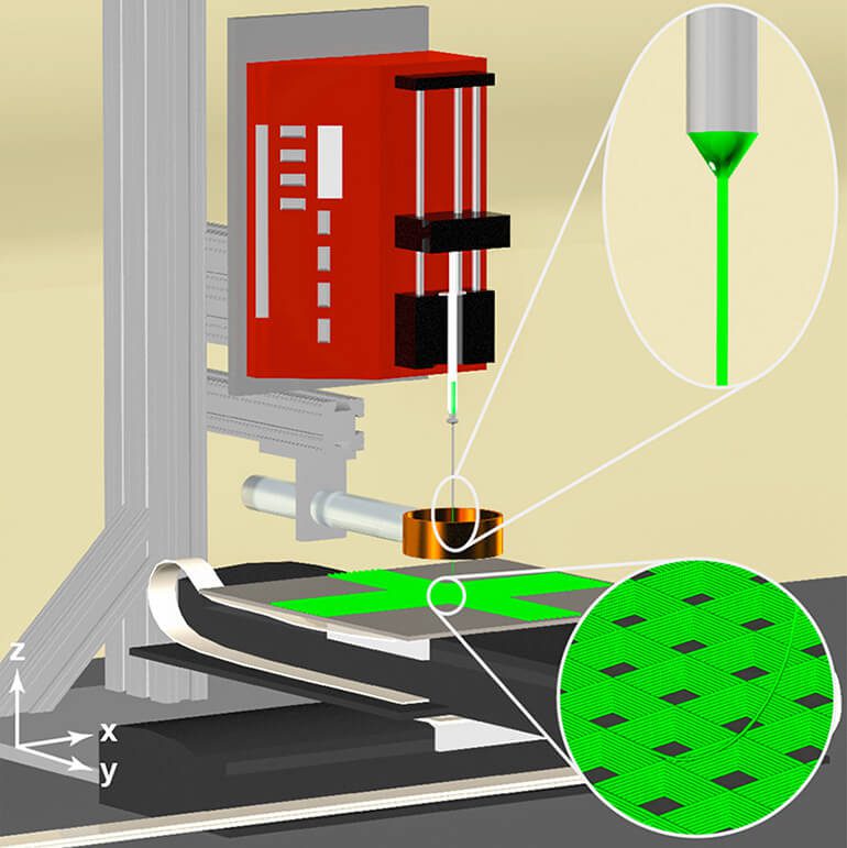
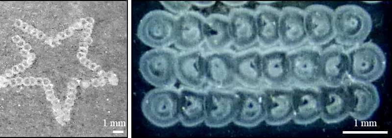
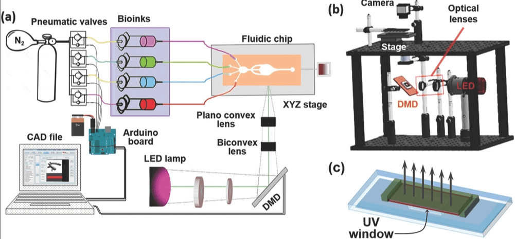
Reading Time: 3 minutesResearchers from UCLA have developed a SLA-based bioprinter that is able to create therapeutic biomaterials from multiple materials. This advancement could potentially be used for on-demand printing of complex artificial tissues for use in transplants and other surgeries. Body tissues are highly complex and made of various different cell types, and this makes it a notoriously […]