Researchers from Purdue University and University of Michigan have developed a new 3D jet writer that allows them to print high-resolution polymer as microtissues. These tiny tissue structures are able to facilitate cancer cell growth, allowing for improved drug development and testing.
A team of researchers at Purdue University and University of Michigan have developed a 3D jet writer that can print high-resolution polymer as microtissues. Since the device is capable of 3D printing on an extremely small scale, the team is able to accurately model pore sizes and recreate a lifelike cancerous environment.
This breakthrough could offer significant opportunities for drug development and testing. Led by Luis Solorio, an assistant professor of biomedical engineering, the research team is aiming to provide better insight into how certain drugs could prevent cancer growth. Moreover, these structures can also improve our understanding of how cancer cells spread throughout the body.
Various researchers have previously experimented with 3D printing to create materials and structures that mimic biological tissues. However, few have been able to achieve the correct porosity to nurture cancer cells to grow and ultimately thrive.

3D Jet Writer Produces Small-Scale Polymer Structures That Mimic Cancerous Environments
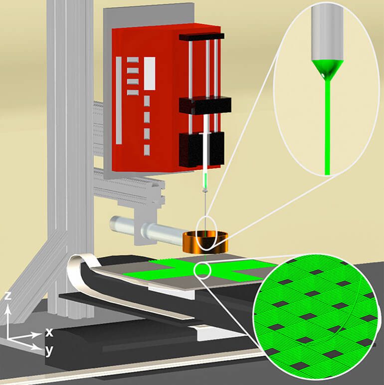
Essentially, 3D jet writing is a more evolved version of electrospinning. With electrospinning, the technique uses a charged syringe with a polymer solution to draw out fiber. Subsequently, researchers are able to arrange the fiber to form a scaffold that enables cell growth.
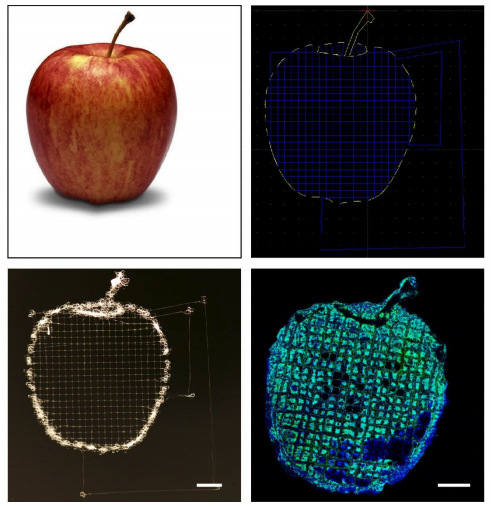
The 3D jet writer developed by the researchers acts similarly to a 3D printer, creating micro tissues from a polymer. However, it does so on a much smaller scale, mimicking the size of pores more effectively. In return, cancer cells can wrap around the structure and grow the same way as they would within a real body.
The team has already tested the viability of these polymer structures in mice. They were able to encourage cancer cell growth in tissues of the subjects, even in areas where cancer would not normally develop. In essence, the experiment demonstrates that the polymer scaffold provides a viable environment for cells to grow.
In the future, the researchers hope that they can utilize the new technique to develop and screen anti-cancer drugs more effectively.
“Ideally, we could use our system as an unbiased drug screening platform where we could screen thousands of compounds, hopefully get data within a week, and get it back to a clinician so that it’s all within a relevant time frame,” states Solorio.
The research paper, entitled “3D Jet Writing: Functional Microtissues Based on Tessellated Scaffold Architectures”, was recently published in Advanced Materials.

Source: Purdue University
Website: LINK
Schreibe einen Kommentar
Du musst angemeldet sein, um einen Kommentar abzugeben.